
什么是免疫检查点(Immune checkpoint)
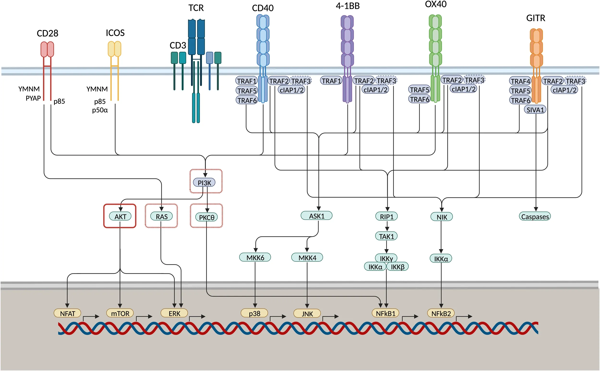
免疫检查点(Immune Checkpoint,IC)是一类在免疫细胞上表达、能调节免疫激活程度的关键因子。它们能够使免疫系统的活化保持在适当范围之内,在有效抵御病原体入侵的同时,防止过度的免疫防御反应和自身免疫性疾病的发生。
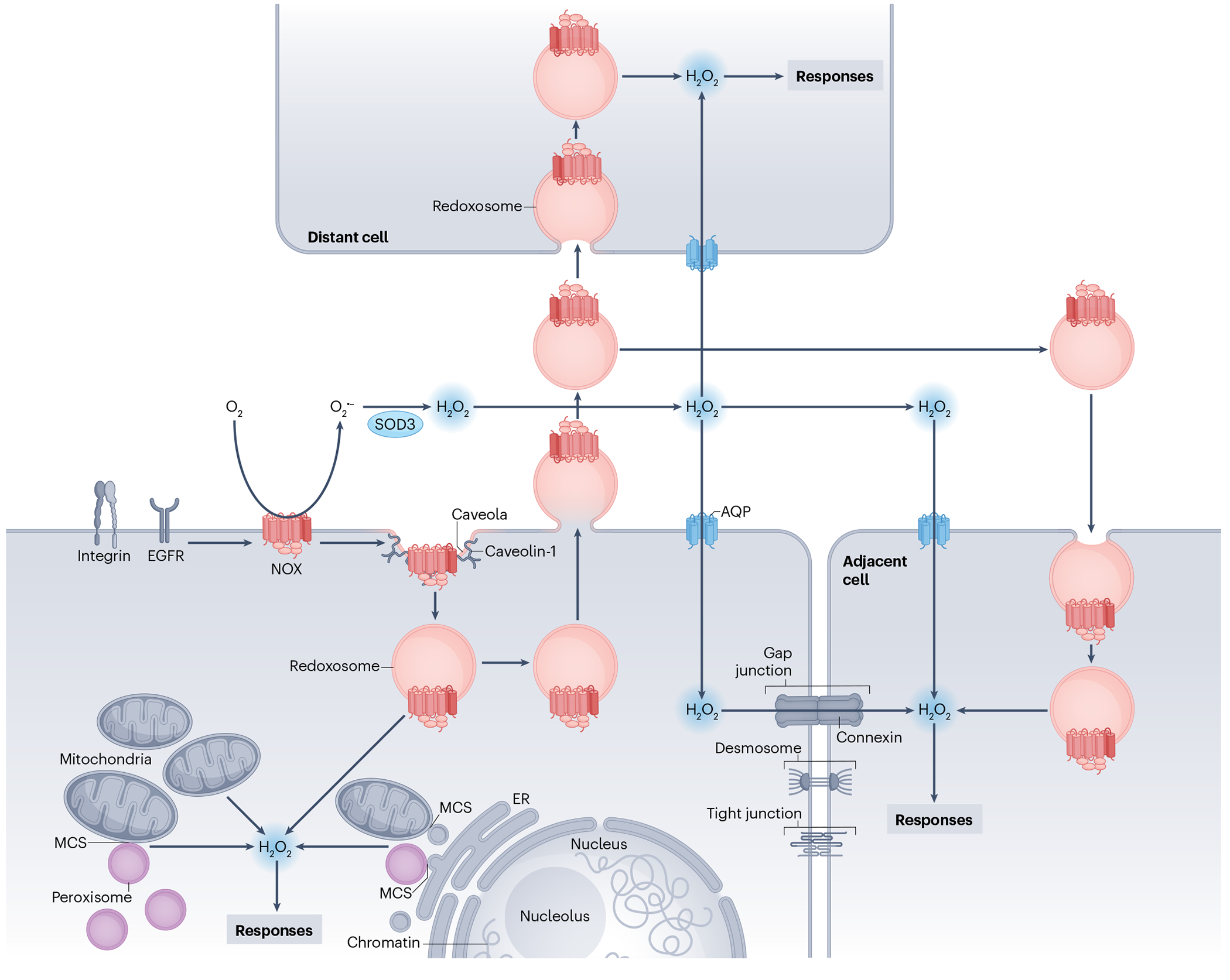
Redox homeostasis denotes the maintenance of the balance between oxidants and antioxidants. Although excessive oxidant production causes oxidative damage to cellular biomolecules and results in oxidative distress, lack of adequate oxidant levels impairs crucial signalling processes and results in reductive distress.
AMPK is a heterotrimeric complex composed of a catalytic α subunit and regulatory β and γ subunits, each of which is encoded by two or three distinct genes (α1, 2; β1, 2; γ1, 2, 3). The kinase is activated by an elevated AMP/ATP ratio due to cellular and environmental stress, such as heat shock, hypoxia, and ischemia. The tumor suppressor LKB1 phosphorylates AMPKα at Thr172 in the activation loop, and this phosphorylation is required for AMPK activation. Accumulating evidence indicates that AMPK not only regulates the metabolism of fatty acids and glycogen, but also modulates protein synthesis and cell growth through EF2 and TSC2/mTOR pathways, as well as blood flow via eNOS/nNOS.
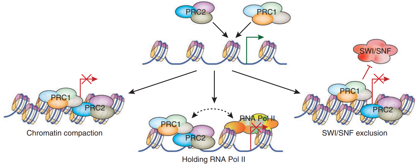
The polycomb group (PcG) proteins contribute to the maintenance of cell identity, stem cell self-renewal, cell-cycle regulation, and oncogenesis by maintaining the silenced state of genes that promote cell lineage specification, cell death, and cell-cycle arrest. PcG proteins exist in two complexes that cooperate to maintain long-term gene silencing through epigenetic chromatin modifications. The first complex, Eed-Ezh2, is recruited to genes by DNA-binding transcription factors and methylates histone H3 on Lys27. This histone methyltransferase activity requires the Ezh2, Eed, and Suz12 subunits of the complex. Methylation of Lys27 facilitates the recruitment of the second complex, PRC1, which ubiquitinates histone H2A on Lys119. PRC1 is composed of Bmi1 and RING1A (also RING1 or RNF1), both of which act to enhance the E3 ubiquitin ligase activity of an additional catalytic subunit RING1B (also RING2 or RNF2). PcG proteins play an important role in the regulation of cell proliferation and senescence through repression of the p16 INK4A and p19 ARF genes and are required for maintenance of adult hematopoietic and neural stem cells, as well as embryonic stem cells.
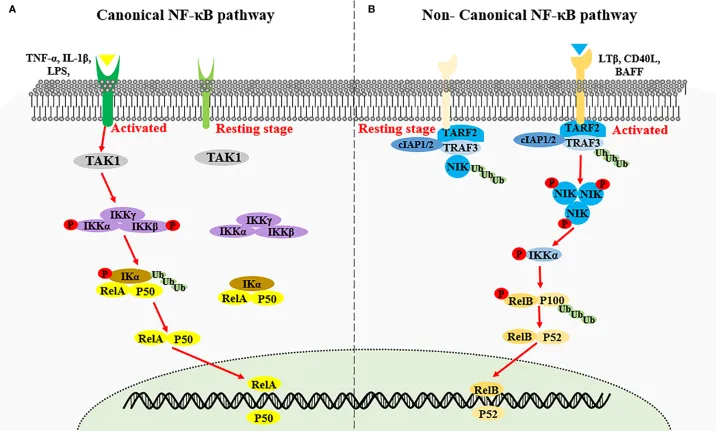
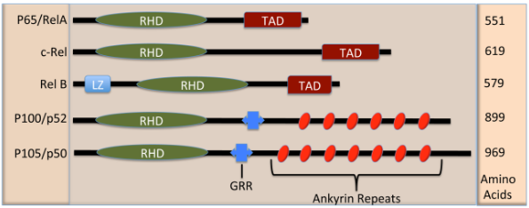
This article elaborates on the activation mechanism, phosphorylation modification of key proteins and transcriptional regulation of the NF-κB signaling pathway. In the cytoplasm, NF-κB remains inactive by binding to inhibitory IκB proteins. The IKK complex phosphorylates IκBα, IκBβ and IκBε, triggering their ubiquitination and proteasomal degradation. The liberated NF-κB dimers then translocate into the nucleus to execute transcriptional functions. The mammalian NF-κB family consists of five members: p65/RelA, c-Rel, RelB, NF-κB1 and NF-κB2. After proteolytic processing, precursor proteins form dimers with Rel proteins to bind DNA. Multiple phosphorylation sites modulate the transcriptional activity of p65. This paper also lists commercial antibodies for research on this pathway with their species reactivity and applicable experimental techniques, along with relevant references, providing a reference for basic research and experimental detection of the NF-κB pathway.
The nuclear factor κB (NF-κB) family participates extensively in inflammatory and immune responses. Distinct from the canonical NF-κB pathway activated by TNFα and LPS, the non-canonical pathway is triggered by BAFF, CD40L and other stimuli. This pathway relies on NF-κB-inducing kinase (NIK) and IKKα to mediate the phosphorylation and partial proteasomal degradation of NF-κB2 p100 into active p52. TRAF2 and TRAF3 act as negative regulators in this process. The non-canonical NF-κB pathway is essential for B cell maturation, the formation of peripheral lymphoid tissues and the prevention of autoimmune diseases. This article also lists a series of specific antibodies for related protein detection, along with relevant research references.
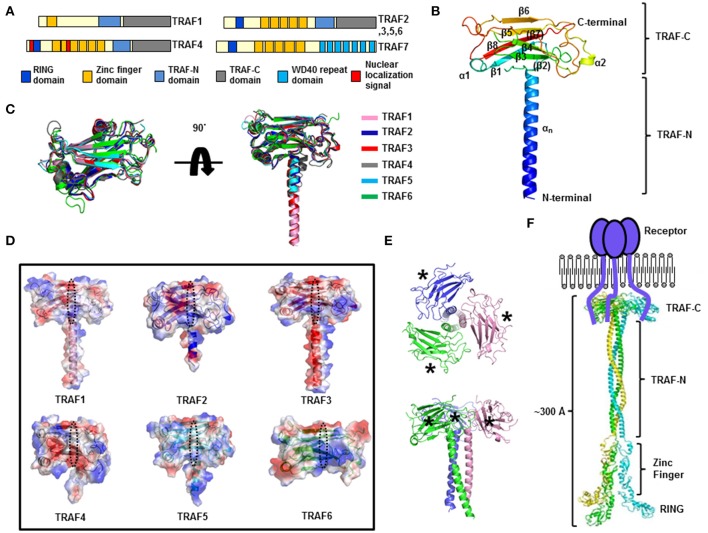
TRAFs (TNF receptor-associated factors) are a family of multifunctional adaptor proteins that bind to surface receptors and recruit additional proteins to form multiprotein signaling complexes capable of promoting cellular responses. Members of the TRAF family share a common carboxy-terminal "TRAF domain", which mediates interactions with associated proteins; many also contain amino-terminal Zinc/RING finger motifs. The first TRAFs identified, TRAF1 and TRAF2, were found by virtue of their interactions with the cytoplasmic domain of TNF-receptor 2 (TNFRII). The six known TRAFs (TRAF1-6) act as adaptor proteins for a wide range of cell surface receptors and participate in the regulation of cell survival, proliferation, differentiation, and stress responses.
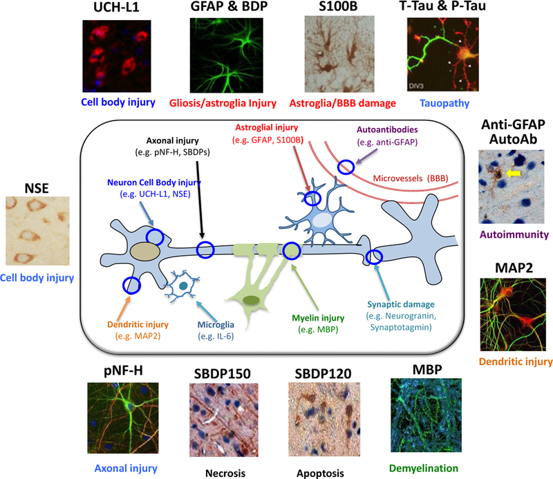
Traumatic brain injury (TBI) is a worldwide health issue that significantly affects the patient as well as their family. Annual total cost of nonfatal TBI in 2016 was $40.6 billion in the United States. In addition to acute brain injury, even mild cases, can lead to cognitive impairment and long-term psychiatric changes. More long term, TBI patients exhibit lower resilience to neurodegenerative disease-associated pathology. Treatment of TBI is made more difficult due to lack of reliable biomarkers to detect TBI. Several proteins are of interest, which are candidates for measurement in blood after TBI. Glial fibrillary acidic protein (GFAP) is an astrocytic intermediate filament protein. As a cytoskeletal protein, GFAP helps provide structural support to astrocytes, which provide metabolic support to neurons and maintains the blood brain barrier. The number and size of astrocytes, in a process called astrogliosis, is also positively correlated with brain injury. Also abundantly expressed in astrocytes, S100B is commonly used as an astrocytic marker and is positively correlated with TBI. Neurofilament-L (NfL) and tau are part of the neuronal cytoskeleton that provide structure to axons. Axons are covered by a multi-layered membrane called the myelin sheath. Myelin basic protein (MBP) is enriched in myelin and helps maintain its structure. UCHL1 and Enolase-2 are ubiquitin hydrolases and glycolytic enzymes, respectively, that are enriched in neurons. PSD95 is an adaptor protein enriched at postsynaptic sites in neurons. After brain injury, neuron-enriched proteins, as well as proteins that maintain neuronal/axonal integrity, can be measured in the blood, reflecting neuronal damage.
The tricarboxylic acid (TCA) cycle includes various enzymatic reactions that constitute a key part of cellular aerobic respiration. The transport of the glycolytic end product pyruvate into mitochondria and the decarboxylation of pyruvate in the TCA cycle generate energy through oxidative phosphorylation under aerobic conditions. Two inner mitochondrial membrane proteins, mitochondrial pyruvate carrier 1 (MPC1) and mitochondrial pyruvate carrier 2 (MPC2), form a 150 kDa complex and are essential proteins in the facilitated transport of pyruvate into mitochondria . Citrate synthase catalyzes the first and rate-limiting reaction of the TCA cycle. Mitochondrial aconitase 2 (ACO2) catalyzes the conversion of citrate to isocitrate via cis-aconitate. IDH1 and IDH2 are two of the three isocitrate dehydrogenases that catalyze oxidative decarboxylation of isocitrate to α-ketoglutarate (α-KG). IDH1 functions as a tumor suppressor in the cytoplasm and peroxisomes, whereas IDH2 is in mitochondria and is involved in the TCA cycle. Mutations in IDH2 have also been identified in malignant gliomas. Dihydrolipoamide succinyltransferase (DLST) is a subunit of the α-ketoglutarate dehydrogenase complex, a key enzymatic complex in the TCA cycle. Succinate dehydrogenase subunit A (SDHA) is a component of the TCA cycle and the electron transport chain and is involved in the oxidation of succinate. Fumarase catalyzes the conversion of fumarate to malate. Fumarase deficiency leads to the accumulation of fumarate, an oncometabolite that has been shown to promote epithelial-to-mesenchymal-transition (EMT), a developmental process that has been implicated in oncogenesis.