
少突胶质前体细胞与少突胶质细胞标志物
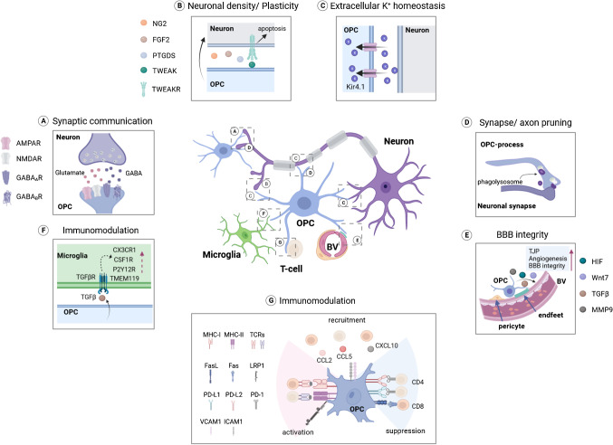
It is now considered that OPCs as neuroectoderm-origin multipotent progenitors that are NG2/PDGFRα positive, highly ramified, mainly generating OLs, but are also capable of giving rise to other CNS cell types including astrocytes.
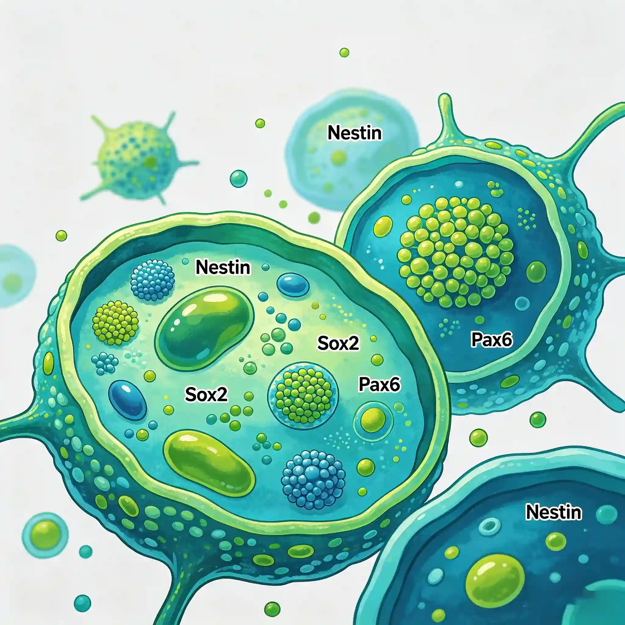
Neuroectodermal markers are a class of biomolecules that are specifically expressed in cells derived from the embryonic neuroectoderm, including proteins, enzymes, receptors, and transcription factors. They are primarily expressed in neurons, glial cells, neuroendocrine cells, neural crest cells, and their corresponding tumor cells.
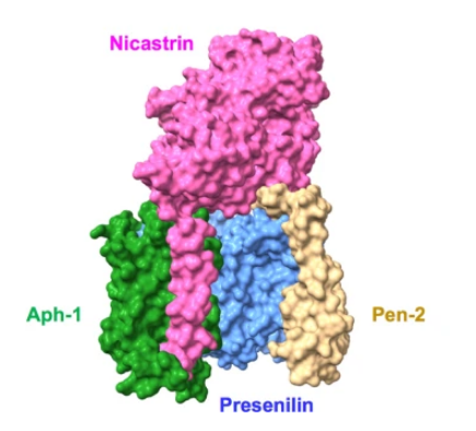
The γ secretase protease complex interacts with and cleaves intramembrane substrates as an essential function for regulation of intracellular signaling and cell-cell interactions. This multiprotein complex is comprised of four integral membrane proteins, Presenilin, Nicastrin, Aph-1, and PEN2, all of which are essential for complete proteolytic activity (1). Presenilin 1 and presenilin 2 are transmembrane proteins belonging to the presenilin family.
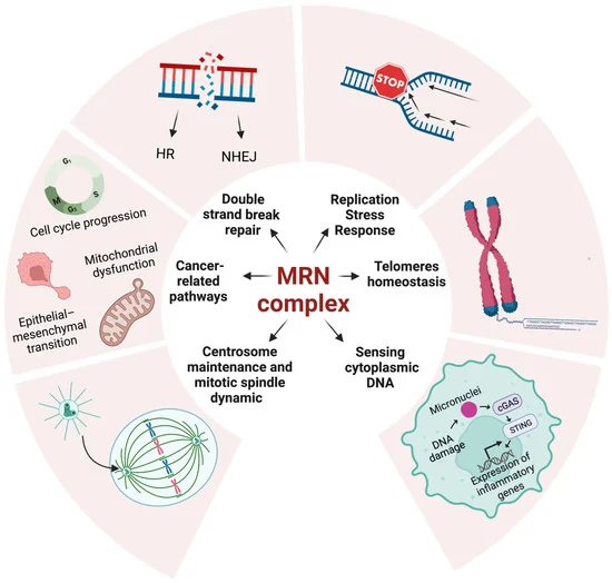
The Mre11-Rad50-Nbs1 (MRN) complex is a critical sensor and regulator in DNA double-strand break repair and genome stability maintenance. It participates in DNA damage repair pathways, checkpoint signaling, and ATM activation, and is closely related to embryonic development, immune cell maturation, and human genetic diseases. This paper introduces the molecular functions of the MRN complex and provides specific antibodies targeting its key components for related experimental research.
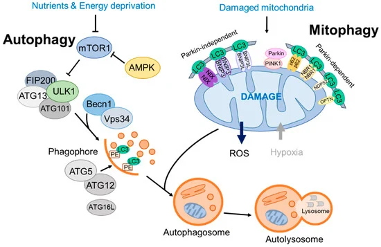
Mitophagy, a pivotal form of selective autophagy, is responsible for the specific elimination of damaged mitochondria to maintain mitochondrial homeostasis and cellular physiological functions. It is regulated by multiple conservative signaling pathways, including the FUNDC1 pathway, BNIP3/BNIP3L pathway, and PINK1/Parkin pathway, which are involved in various physiological and pathological processes such as hypoxia adaptation, erythroid cell maturation, and neurodegenerative diseases. This paper systematically elaborates on the core molecular mechanisms of mitophagy and collates a panel of high-quality antibodies targeting key proteins of mitophagy (e.g., LC3B, PINK1, Parkin, p62/SQSTM1, BNIP3). These antibodies are applicable to multiple experimental techniques including Western Blot, immunofluorescence, immunohistochemistry and immunoprecipitation, providing reliable molecular tools for the in-depth study of mitophagy-related biological processes and disease mechanisms.
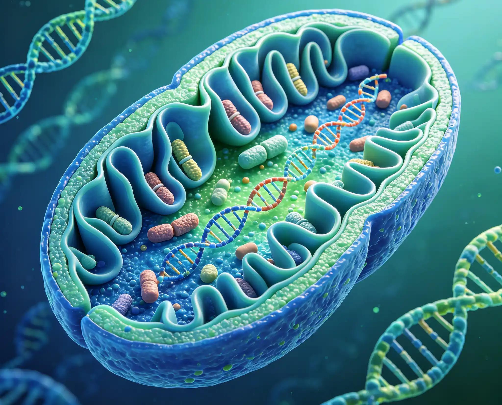
This study focuses on the Mitochondrial Marker Antibody Sampler Kit, which contains a variety of antibodies targeting classic mitochondrial proteins, including key mitochondrial proteins such as cytochrome c oxidase, cytochrome c, HSP60, prohibitin, pyruvate dehydrogenase complex, succinate dehydrogenase, SOD1, and voltage-dependent anion channel. These proteins are located in different substructures such as the inner mitochondrial membrane, intermembrane space, matrix, and outer membrane, and participate in core life activities including mitochondrial electron transport chain, tricarboxylic acid cycle, protein folding and stability, and metabolite transport. This paper systematically presents the product information of 8 rabbit monoclonal antibodies targeting the above mitochondrial proteins, as well as 2 matched secondary antibodies and internal reference antibodies, including catalog numbers, reactive species, and applicable experimental types.
The nucleosome, made up of four core histone proteins (H2A, H2B, H3, and H4), is the primary building block of chromatin. Originally thought to function as a static scaffold for DNA packaging, histones have now been shown to be dynamic proteins, undergoing multiple types of post-translational modifications, including acetylation, phosphorylation, methylation, and ubiquitination (1). Histone methylation is a major determinant for the formation of active and inactive regions of the genome and is crucial for the proper programming of the genome during development (2,3). Arginine methylation of histones H3 (Arg2, 17, 26) and H4 (Arg3) promotes transcriptional activation and is mediated by a family of protein arginine methyltransferases (PRMTs), including the co-activators PRMT1 and CARM1 (PRMT4) (4). In contrast, a more diverse set of histone lysine methyltransferases has been identified, all but one of which contain a conserved catalytic SET domain originally identified in the Drosophila Su(var)3-9, Enhancer of zeste, and Trithorax proteins.
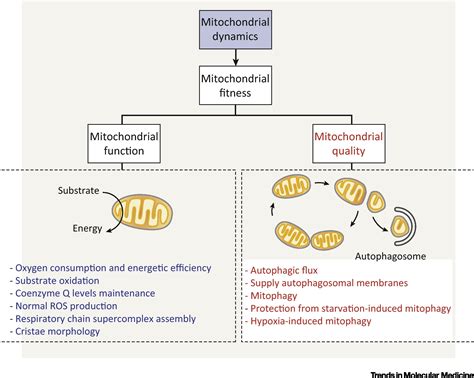
Mitochondrial dynamics, encompassing fission and fusion processes, plays a critical role in the regulation of mitochondrial morphology, metabolism, apoptosis, and autophagy. Protein import into mitochondria is primarily mediated by the translocase of the outer mitochondrial membrane (TOM) complex and the translocase of the inner membrane (TIM) complex, in which Tom20, a key component of the TOM complex, initiates the recognition of protein precursors. Mitochondrial dynamics are tightly governed by dynamin-related GTPases: DRP1 modulates mitochondrial fission, while mitofusin-1, mitofusin-2, and OPA1 control mitochondrial fusion. The activities of these core proteins are precisely regulated via post-translational modifications. OPA1 is regulated by alternative splicing and proteolytic processing, and its expression can be induced by metabolic stress. DRP1 activity is differentially modulated by phosphorylation at distinct sites, with Ser616 phosphorylation promoting fission and Ser637 phosphorylation inhibiting this process. Furthermore, mitochondrial fission factor (MFF) acts as a receptor for DRP1, and its phosphorylation by AMPK facilitates the recruitment of DRP1 to mitochondria. This review summarizes the molecular mechanisms underlying mitochondrial protein import and the regulatory network of mitochondrial dynamics.